
 217 Promenade Way, Unit 302


- Echocardiogram
- Vascular Ultrasound
- General Ultrasound

Unit 302
Sugar Land, Texas 77479
View Map & Directions
Phone: 713-715-7056
roy@precisioncardiovasculardiagno
stics.com
- Venous Reflux Ultrasound
- Ultrasound guidance -Radio frequency thermal & Laser vein ablation procedure (CPT 36475)
- Cardiac Ultrasound
- Vascular Ultrasound
- Carotid Ultrasound
- General Ultrasound
- ABI (Ankle Brachial Index)
- Holter Monitors
All of our registered Sonographers are certified in all 3 ultrasound modalities. Here is a list of ultrasound exams that we can offer to your medical practice:
- Echocardiogram (M-mode, 2D & Doppler)
- Stress Echo (Tread Mill)
- Echo Screenings
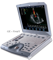 An echocardiogram is a test in which ultrasound is used to examine the heart. In addition to providing single-dimension images, known as M-mode echo that allows accurate measurement of the heart chambers, the echocardiogram also offers far more sophisticated and advanced imaging. This is known as two- dimensional (2-D) Echo and is capable of displaying a cross-sectional "slice" of the beating heart, including the chambers, valves and the major blood vessels that exit from the left and right ventricle.
- Intima Media Thickness (IMT)
- Venous Doppler - Lower & Upper (R/O DVT)
- Arterial Doppler - Lower & Upper (R/O PAD)
- ABI - Ankle Brachial Index (Arterial circulation)
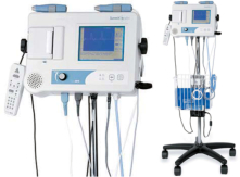
Vascular ultrasound provides pictures of the body's veins and arteries physiological and pathological conditions. Vascular Doppler studies enable to detect DVT's (Deep Vein Thrombosis) & PAD (Peripheral Arterial Disease) and individuals who are high risk for stroke. Doppler ultrasound is a special ultrasound technique that evaluates blood velocity as it flows through a blood vessel, including the body's major arteries and veins in the abdomen, arms, legs and neck.
Carotid ultrasound is a test that shows the carotid arteries (vessels in the neck that provide blood flow to the brain), as well as how much blood flows and how fast it travels through them. Ultrasound waves - the same ones used in imaging the fetus in a pregnant woman - are used to make an image of the arteries. This image can be used to find out if there is an abnormality or blockage of the carotid arteries that could lead to stroke. This test can be used to investigate the carotid arteries for several reasons, but the information here applies only to stroke evaluation.
- Renal Artery Doppler
- Testicular Ultrasound
- Retroperitoneal (Kidney & Aorta) Ultrasound
- Thyroid Ultrasound
- Abdominal Aorta Ultrasound
- Bladder Ultrasound
- Breast Ultrasound
- Abdominal Ultrasound
- Spleen
- Liver
- Gallbladder
- Pancreas
- Bladder
- Pelvis
An ankle brachial index, or ABI, is performed to check for peripheral artery disease (PAD). Peripheral artery disease is a condition involving blockages in the arteries of the arms or legs. If left untreated, peripheral artery disease can cause leg pain, stroke, heart attack, or poor circulation.
No special preparations are necessary before undergoing an ankle brachial index. During the ankle brachial index test, you will lie down on a table, and a technician will measure your blood pressure in both arms. The technician will then measure your blood pressure in arteries in the left ankle and use a Doppler ultrasound to produce images of those arteries. An ankle brachial index usually only takes a few minutes and is painless. This test is done by measuring blood pressure at the ankle and in the arm while a person is at rest. The ABI result can help diagnose peripheral arterial disease (PAD). A lower ABI means you might have PAD.
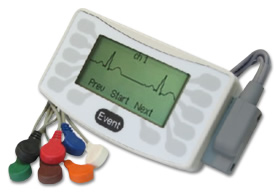
A Holter monitor is a continuous tape recording of a patient's EKG for 24 hours. Since it can be worn during the patient's regular daily activities, it helps the physician correlate symptoms of dizziness, palpitations (a sensation of fast or irregular heart rhythm) or black outs. Since the recording covers 24 hours, on a continuous basis, Holter monitoring is much more likely to detect an abnormal heart rhythm when compared to the EKG which lasts less than a minute. It can also help evaluate the patient's EKG during episodes of chest pain, during which time there may be telltale changes to suggest ischemia or reduced blood supply to the muscle of the left ventricle.